Developmental Dysplasia of the Hip (DDH) â general Information
Introduction
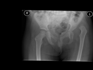
Developmental Dysplasia of the Hip (also known as congenital dislocation of the hip) is a variable condition. It occurs when the hip fails to grow properly in the socket. The socket becomes shallow and the hip can eventually dislocate from the joint. The condition can present from birth to adulthood. The condition varies between a slightly shallow hip to a completely dislocated hip joint. In babies and young children, the condition is not painful and does not affect movements of the leg or walking ability. The exact cause of the condition is unknown. However it is more common in girls than boys, itâs more common in first-born children and it is more common if a first degree relative is affected by the condition.
What happens if DDH is not treated?
Untreated DDH can lead to a limp because of a short leg. It does not cause pain in young children. It does not lead to delayed walking. In teenage or young adult life, hip and back pain can occur. The hip can develop early onset arthritis.
How can DDH be treated?
The aim of treating the condition is to reduce the ball part of the hip back into the socket and keep it in place. This then allows the hip to continue growing as normally as possible. The hip should be reduced gently without any tension to reduce the chance of further growth problems of the hip.
Treatment of this condition varies with the degree of dislocation of the hip and the age of the child.  I will discuss the treatment that is required. The different treatment options are described below.
 Pavlik Harness Treatment
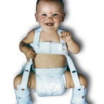
The Pavlik Harness is used to treat Developmental Dysplasia of the Hip in children upto the age of 6 months. It consists of a fabric harness which helps to hold the hips flexed and out to the side. It is used to help guide the hips back into sockets and to encourage them to grow. A baby is allowed to kick their legs in the harness and can be picked up and nursed in the normal manner. The harness normally has to stay on for 24 hours a day, but later on during treatment can be removed for bath times. Treatment is monitored using regular ultrasound scans. Your child will be brought to a special clinic where the harness will be checked, the child examined and the scan undertaken.
The Period of treatment varies depending on the condition of the hip but it is normally in the order of 3 to 4 months. Further follow up is required long term to assess the growth of the hip. This treatment method has a high success rate of over 90%.
Complications of Treatment
In a small percentage of children, the Pavlik Harness does not hold the hip in joint and further treatment is required. In a small percentage of children the blood supply to the hip can be affected, resulting in a growth disturbance of the hip (avascular necrosis).
 Closed Reduction and Hip Spica.
This is a method used to treat Developmental Dysplasia of the Hip in children from the age of 3 months up until 12 months. It is used where Pavlik Harness treatment has failed or where the child presents at a later age. The aim of treatment is to allow the hip to go back into socket and to hold it in a body plaster ( hip spica)
This treatment involves bringing your child into hospital the day of the procedure. The child is given a general anaesthetic so they are asleep and relaxed. The hip is then examined and a small needle is put into the hip joint to inject some dye. This outlines the structures within the hip when x-rays are taken. Occasionally a small tight tendon in the groin needs to be released during this procedure to improve the movement of the hip. This is a small cut in the groin approximately 0.5 cm long. If the hip reduces without any tension and is stable, then it is held in place by a hip spica. This procedure takes approximately 45 minutes.
The child normally recovers quickly from this procedure. The nurses on Alice Ward will advise you about how to look after your child in the spica. A special scan (MRI scan) is taken of the hip 2 days after to check that the hip has remained in place. The total time in hospital is between 4 and 5 days. The spica is kept on for a total of 4 months. However it is changed under a brief anaesthetic after 2 months to allow for growth of the child and for hygiene. This second procedure can be performed as a daycase. After 4 months in the spica, the child is brought back to the ward and the plaster is removed without anaesthetic. A plastic splint is applied to help hold the legs apart. The child will also have some physiotherapy to help move the joints. This normally requires a stay in hospital of 2-3 days. The plastic splint is worn for a total of 16 weeks. During this time, the child can move around, and older children will be able to crawl and even walk in the plastic splint.
In approximately 10% of cases, closed reduction is not successful because the hip is too tight or unstable. In this case the child is allowed to go home the same day and brought back at a later date for a surgical procedure (See below).
Complications
Complications of this procedure are uncommon. In a small percentage of children, blood supply to the growing hip will be affected (avascular necrosis). The majority of children affected by this condition do not require any further treatment. Occasionally there can be problems with the hip spica. The skin can become irritated or sore underneath. This normally settles on removal of the spica. Following the closed reduction treatment, the child is followed up long term to ensure the hip grows normally. If the hip remains persistently shallow, then occasionally further surgery is required.
 Open Reduction of the Hip
This is an open surgical procedure which allows the hip to go back into socket by releasing tight structures around the hip. It is normally performed on children from the age of 6 months to 2 years. The child is brought in the day of surgery. The operation is performed under a general anaesthetic. An incision is made in the groin. The hip joint is opened, tight structures are released and the hip is put back into joint. The lining of the joint is tightened to hold the hip in place. The wound is closed with stitches which dissolve under the skin. The child is then placed into a body plaster (hip spica) which is kept on for 3 months The total stay in hospital is normally 5-6 days. A special scan (MRI scan) is taken 2 days after the operation to check that the hip has remained in place.
After 3 months, the child is brought back into hospital for the plaster to be removed. The child will need to have physiotherapy in hospital for approximately 1 week to regain mobility. The child will then be followed up in the outpatient clinic at regular intervals to monitor the growth of the hip
Complications
There will be a scar in the groin. This generally fades well, but can occasionally be more obvious.
Whenever there is a cut made in the skin, there is a small chance of infection. Serious infection is rare in children.
There is a chance of injuring nerves and blood vessels around the hip. This is rare.
The blood supply to the growing hip can be affected (avascular necrosis). This is uncommon, and does not always need treatment. Rarely, it can lead to a more serious disturbance to the growth of the hip, and may require further operations.
The hip requires regular follow up with X rays to ensure that the shallow hip socket grows deeper. If this does not occur, then a further procedure can be required
 Femoral Osteotomy and Salterâs Pelvic Osteotomy
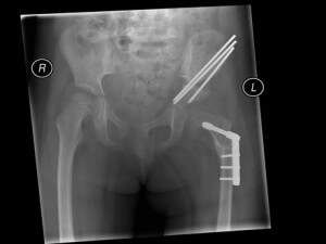
In older children presenting with Developmental Dysplasia of the Hip or following previous treatment for Developmental Dysplasia of the Hip, additional surgery can be required. The open reduction operation can be combined with a femoral and / or a Salterâs pelvic osteotomy. A Femoral Osteotomy means cutting the thigh bone. The thigh bone is then re-aligned, shortened, and held in place with a plate and screws. The femoral osteotomy operation is performed through an incision along the side of the thigh. If there is a shallow hip socket then a Salterâs Pelvic Osteotomy can be undertaken. This is performed through the same incision in the groin as used for an Open Reduction. The pelvic bone is cut and the socket is rotated to help treat shallowness of the hip socket. The socket is held in place with a bone graft taken from the hip bone. It is held in place with two wires. The wires are underneath the skin. For both the Femoral and Salterâs Pelvic Osteotomy, the stitches are below the skin and do not need to be taken out. The child is held in a body plaster (hip spica). This is normally for a period of 6 weeks. For the child having Open Reduction, Femoral Osteotomy and a Salterâs Osteotomy, the stay in hospital is 5-7 days. A CT scan is taken a few days after surgery to check that the hip has remained in place. The child is brought back to the ward 6 weeks after the operation for removal of the plaster. This does not require an anaesthetic. The child does not require any further splintage. Long term follow up is required to monitor the growth of the hip. Approximately 6 months to one year after surgery, the metal pins, plates and screws are removed. This requires a further operation but the child does not have to go into a plaster on this occasion.
Complications
Surgery for a Femoral or Pelvic Osteotomy leaves a scar. The scar on the thigh can be more prominent than the scar in the groin. Scars grow as the child grows. There is a small possibility of injuring nerves and blood vessels in the area of surgery. There is a small possibility of infection though this is rarely a problem in children. For a child having a Femoral Osteotomy, a section of bone is removed to allow the hip to sit in socket without any tension. This results in the operating leg being slightly shorter than the normal leg. This shortening normally corrects itself with growth, but is monitored closely.


